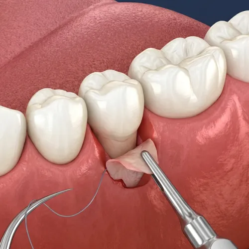
Gum Graft Disadvantages, Risks, and Pain: What Patients in Encino Should Know
Wondering about gum graft risks, pain, and drawbacks? Learn what Encino patients must know before surgery. Schedule your expert consultation with us today!
Read More

Emergency Dentistry
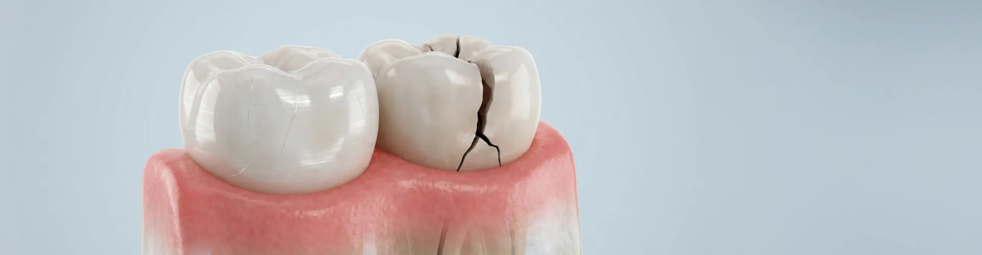
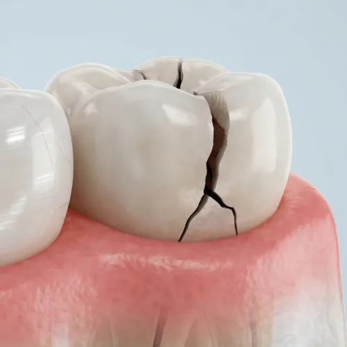
Not every crack is a dental emergency, and not every cracked tooth needs a root canal — but some cracks silently destroy the bone beneath them. The right treatment depends on objective diagnostic tests and imaging that reveal exactly how deep and how far a crack has traveled.
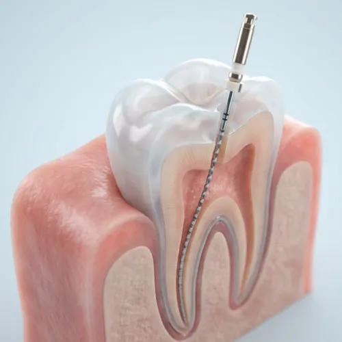
Patients are often surprised to learn that the naked eye — even a trained one — isn't enough to diagnose a cracked tooth accurately. According to the American Association of Endodontists, symptoms like erratic pain during chewing or sudden sensitivity to temperature changes are classic signs, but they don't tell the whole story about crack depth or location. Understanding the parts of a tooth — from enamel and dentin to the pulp and root — helps explain why crack depth matters so much for treatment decisions.
Three specific tools change that:
The Tooth Slooth Bite Test. This small plastic device isolates individual cusps. A patient bites down, then releases. Pain that spikes on release — not on biting down — signals a structural crack flexing and springing back. A craze line (a purely cosmetic surface mark) produces no such response.
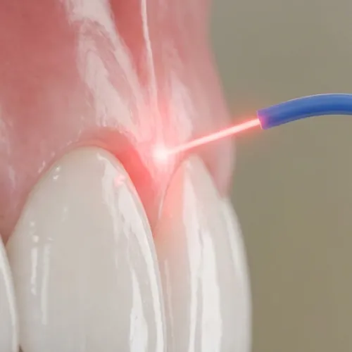
Transillumination. A high-intensity fiber-optic light is passed through the tooth. Light travels freely through a craze line but stops abruptly at a true fracture. The shadow created by that interruption maps the crack's direction and approximate extent. Research published by the NCBI confirms that fiber-optic transillumination, combined with visual magnification, is one of the most reliable front-line diagnostic tools for cracked tooth syndrome.
Dental Dye Staining. A dye is applied to the tooth surface. It seeps into structural cracks but not into craze lines, making the fracture visible under magnification.
Together, these three tests give a dentist objective, reproducible data. If all three are negative, watchful monitoring is reasonable. If any are positive, the crack is structural — and the next question becomes how deep it goes.
This is where many cracked tooth cases go wrong. Standard bitewing and periapical X-rays are excellent at detecting decay and bone loss, but they have a critical blind spot: vertical cracks running along the length of a root are nearly invisible on 2D film.
A patient with a deep crack may receive a root canal and crown — a significant procedure — only for it to fail months later. The crack had already reached the root, and the 2D image simply couldn't show it. The NIH found that bout 32% of patients with an untreated cracked tooth experience a change in symptoms within one year, which underscores how quickly a manageable crack can become a root-level problem.
Cone Beam Computed Tomography (CBCT) solves this. A CBCT scan produces 3D axial cross-sections of the tooth and its root, allowing a specialist to visualize micro-fractures in the canal wall that are simply not visible on any 2D image. Before committing to a root canal on a deeply cracked tooth, CBCT imaging is the standard that separates a treatable crack from one that has already reached the point of no return. Our advanced dental technology makes this level of precision imaging available to patients in Encino.
The NCBI notes that CBCT is "indispensable in cases where conventional imaging methods are insufficient" — precisely the scenario of a suspected deep root crack.
There's a longstanding assumption in dentistry that any crack extending below the gumline is an automatic extraction. The evidence no longer supports that view.
Research highlighted by the American Association of Endodontists shows that endodontically treated cracked teeth — even those with cracks extending onto the root surface — demonstrate survival rates ranging from 82% to 96%. One modified treatment protocol produced over 90% success and 96% survival rates over two to four years, outcomes comparable to root canal treatment on non-cracked teeth.
The key modifiers in that protocol matter practically:
The crown is non-negotiable. A root canal without prompt full-coverage restoration dramatically increases the risk of the crack propagating further. Cleveland Clinic explains that without treatment, a cracked tooth opens the door for bacteria to reach the pulp, causing an abscess that can spread to the jawbone. Understanding the cost of root canal treatment can help patients plan ahead when this procedure becomes necessary.
Our endodontics specialists are trained to apply these precise protocols, giving deeply cracked teeth the best possible chance of long-term survival.
Even with the best endodontic technique, some cracked teeth cannot be saved. The clearest radiographic signal is a pattern called a "J-shaped" or halo radiolucency — a dark shadow on an X-ray that wraps around the root of the tooth like a parenthesis.
This shadow indicates localized bone resorption. Bacteria leaking through the crack have been destroying the bone around the root, often silently and without pain. By the time this pattern appears, the structural damage to the socket is typically irreversible.
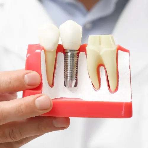
Why does this matter beyond the tooth itself? Patients who plan to replace an extracted tooth with dental implants need adequate bone volume in the socket. A tooth left in place with active J-shaped bone loss continues to erode that volume. Extraction at the right time — before the halo widens — preserves the bone needed for a successful implant later.
A WebMD overview of cracked tooth repair notes that even with treatment, tooth cracks sometimes continue to get bigger or split, which can result in eventual tooth loss. The J-shaped pattern is the point at which continuing to treat the tooth stops benefiting the patient and starts depleting the bone they'll need for the next phase of care.
If you're dealing with tooth pain on biting, unexplained sensitivity, or a crack your general dentist hasn't fully assessed, Akemi Dental Specialists offers comprehensive cracked tooth evaluation in Encino, California. Our team uses advanced diagnostic protocols — including bite testing, transillumination, and 3D imaging — to give you a clear, evidence-based answer about your treatment options before any procedure is recommended. Learn about habits and pro tips to maintain your dental implants if implant placement becomes part of your treatment plan. Contact us to schedule your consultation.
This article is intended for informational purposes only and does not constitute professional dental or medical advice. Always consult a licensed dental professional for diagnosis and treatment recommendations specific to your situation.


Dental Specialists Encino, CA
Here's what our patients are saying about us...





Mon, Fri 6:30 am - 4 pm
Tue, Thu 6:30 am - 5 pm
Wed 6:30 am - 3 pm
Sat, Sun Closed
16055 Ventura Blvd. Suite #820, Encino, CA 91436
Endodontic Care Encino, CA